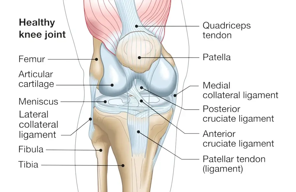
At SIGMA Orthopedics & Sports Medicine, we use advanced imaging, high-definition arthroscopy, and a tissue-preserving surgical philosophy to treat meniscus injuries with unmatched accuracy.
Our mission: identify the exact tear pattern, protect your knee for the long term, and get you back to peak performance.
Lorem ipsum dolor sit amet, consectetur adipiscing elit. Ut elit tellus, luctus nec ullamcorper mattis, pulvinar dapibus leo.
Every evaluation and procedure follows rigorous, high-reliability surgical pathways designed to reduce variability and increase safety.
Where many surgeons remove damaged tissue, SIGMA prioritizes repair over removal whenever possible — protecting your knee for decades.
Our advanced diagnostic workflow integrates specialized MRI interpretation and high-definition 4K/8K arthroscopy for unmatched clarity.
Your recovery begins on day one — with a structured, measurable rehab pathway engineered for athletes, executives, and active adults.
he meniscus is a C-shaped cartilage cushion that absorbs shock, stabilizes the knee, and protects the joint from wear.
Each knee has two menisci — medial (inside) and lateral (outside).
A meniscus tear occurs when this cartilage is damaged from twisting, squatting, sudden rotation, or direct impact. Tears can be:
At SIGMA, identifying the exact tear pattern matters — because it determines whether the meniscus can be repaired or must be trimmed.

Look for the following symptoms — especially after a twist, pivot, or sudden injury:
If these sound familiar, you may need a definitive evaluation — especially if symptoms persist for more than 2–4 weeks.

Appropriate for stable tears or degenerative patterns:
We reassess after a short recovery window to ensure progress.
We use sutures and anchors to re-approximate the tear edges, allowing the tissue to heal naturally.
Best for:
Benefits:
To improve healing, we may add platelet-rich plasma or bone marrow concentrates during repair — especially important for borderline tear patterns.
If the tear is in a zone with no healing potential, we selectively remove only the damaged fragment — protecting all remaining healthy meniscus.
SIGMA’s approach: trim as little as possible to preserve your long-term joint health.
Strength, mobility, nutrition, and swelling management — planned using Six Sigma risk-reduction methods
Performed through tiny incisions using high-definition optics and specialized instruments.
Guided exercises begin immediately, preventing stiffness and protecting the repair.
A comprehensive, progressive rehab plan designed for athletes and active adults.
You will undergo functional, strength, and biomechanical testing before full release.
Advanced Physical Examination
We assess joint line tenderness, stability, motion patterns, and mechanical symptoms.
High-Resolution MRI Review
We look beyond standard reports — identifying subtle tear configurations often overlooked.
Dynamic Diagnostic Arthroscopy (when needed)
If pain persists despite “normal” imaging, high-definition arthroscopy can directly confirm the diagnosis and guide same-day treatment.
All procedures carry risk — but at SIGMA, risk reduction is engineered into every step.
Potential risks:
How we minimize risk:

We continuously adopt new technologies to improve meniscus outcomes:
SIGMA’s vision: a future where meniscus preservation is the standard — not the exception.

At SIGMA Orthopedics & Sports Medicine, you’ll receive:

For more information on meniscus tears of the knee, or the excellent treatment options available, please contact the office of Frank McCormick, MD, orthopedic knee specialist serving Orlando, West Palm Beach County, and surrounding Florida communities.